
Pupil evaluation is a critical component of neurological assessments, providing valuable insights into a patient’s neurophysiological state. Pupil diameter measurement, or pupillary size measurement, can reveal a range of neurological conditions and help clinicians make informed decisions. This blog will guide you through interpreting pupil evaluation findings using advanced neurological tools and the Neurological Pupil Index (NPi).
Understanding Pupil Evaluation
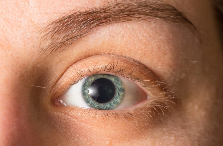
Pupil evaluation involves measuring the size, shape, and reactivity of the pupils to light and other stimuli. It is a fundamental aspect of a neuro exam, offering an immediate glimpse into the functionality of the cranial nerves, particularly the optic nerve (CN II) and the oculomotor nerve (CN III). Variations in pupil size and reaction can indicate issues such as increased intracranial pressure, brain injury, or optic nerve dysfunction.
The Importance of Pupil Diameter Measurement
Pupil diameter measurement is essential in detecting and diagnosing various neurological disorders. The size of the pupils can change due to several factors, including light exposure, stress, and certain medications. However, when these changes are asymmetrical or abnormal, they can indicate serious underlying conditions.
For instance, anisocoria (unequal pupil sizes) may suggest Horner’s syndrome, third cranial nerve palsy, or a brain aneurysm. By accurately measuring pupillary size, healthcare professionals can identify these conditions early and initiate appropriate interventions.
Interpreting Findings with the Neurological Pupil Index (NPi)
The Neurological Pupil Index (NPi) is a calculated score that offers a standardized way to interpret pupil evaluation findings. It is derived from various parameters, including pupil size, reactivity, and latency, measured using advanced pupillometry devices. The NPi provides an objective metric to assess a patient’s neurological status, with a normal score typically ranging between 3 and 4.
An NPi score below 3 may indicate abnormal pupillary function, potentially signaling increased intracranial pressure or brainstem dysfunction. Clinicians rely on the NPi for continuous monitoring of patients with traumatic brain injuries, strokes, or other conditions that could lead to neurological deterioration. By tracking changes in the NPi, healthcare providers can detect early signs of complications and adjust treatment plans accordingly.
Tools for Accurate Pupil Measurement
The accuracy of pupil evaluation depends heavily on the tools used during the assessment. Traditional methods involve using a penlight and a ruler, which can be subjective and prone to human error. However, advanced neurological tools have revolutionized this process, offering precise and reliable pupillary size measurements.
One such tool is the automated pupillometer, which uses infrared technology to measure pupil size and reactivity objectively. These devices provide standardized measurements, eliminating the variability associated with manual methods. The use of pupillometers is especially critical in intensive care units (ICUs) and emergency departments, where rapid and accurate assessments are vital.
Clinical Applications of Pupil Evaluation
Pupil evaluation plays a critical role in various clinical settings, particularly in neurology, neurosurgery, and critical care. In patients with head trauma, for example, pupil evaluation is one of the first assessments performed. A dilated or non-reactive pupil could suggest a brain herniation, necessitating immediate medical intervention.
In stroke management, pupil evaluation can help differentiate between ischemic and hemorrhagic strokes. Abnormal pupil findings may indicate bleeding in the brain, which requires different treatment approaches compared to ischemic strokes.
Furthermore, pupil evaluation is essential in monitoring the effects of certain medications. For example, opioid use can cause pinpoint pupils, while atropine can lead to dilated pupils. Understanding these pharmacological effects is crucial for the accurate interpretation of pupil evaluation findings.
Conclusion
Pupil evaluation is one of the most powerful neurological tools in neurological assessments, providing vital information about a patient’s brain function and overall neurological status. Accurate pupil diameter measurement and interpretation of findings through advanced tools like the NPi can significantly enhance clinical decision-making. As technology continues to evolve, the integration of automated pupillometers and the use of standardized metrics like the NPi will further improve the accuracy and reliability of pupil evaluations, ultimately leading to better patient outcomes.